Oncogenes and Oncoproteins


Antibodies for the Analysis and Characterization of Oncoproteins

Mutations in growth factors, growth factor receptors, cell cycle regulators, cell signaling proteins and transcription factors tend to result in dominantly active oncogenes, which alter the balance between cell proliferation and apoptosis.
Cancer is a complex disease that can be triggered by a variety of causes ranging from oncovirus infections to genetic mutations. Exposure to carcinogens and inheritance are two of the main sources for genetic mutations and predominantly occur in seven distinct protein groups (Lodish et al. 2000):
- Apoptosis regulators
- Cell cycle control proteins
- Cell signaling proteins
- DNA repair proteins
- Growth factors
- Growth factor receptors
- Transcription Factors
Mutations in growth factors, growth factor receptors, cell signaling proteins and transcription factors tend to result in dominantly active oncogenes; a term used to describe genes, which encode a cancer-promoting oncoprotein. Most oncogenes are activated via point mutations, gene amplifications and gene translocations in a normal gene known as proto-oncogene and result in altered gene expression or protein activity levels (Lodish et al. 2000).
Bio-Rad offers a variety of antibodies and proteins for the analysis of oncoproteins by ELISA, Flow Cytometry, Immunofluorescence, Immunohistochemistry and Western Blotting.
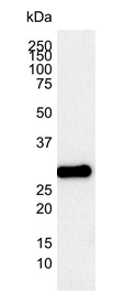
Western blot analysis of a Jurkat cell lysate with mouse anti-human Bcl-2 antibody (MCA1550) followed by detection with a goat anti-mouse HRP conjugated secondary antibody. Visualization was performed by Chemiluminescence.
Featured Murine Anti-Bcl-2 Antibody (MCA1550)
The apoptosis regulator Bcl-2 oncoprotein plays an important role in the decision of whether a cell lives or dies. It acts by controlling the permeability of the outer mitochondrial membrane.
Murine anti-Bcl-2 antibody (clone 100) is frequently used in Immunohistochemistry experiments and has been successfully used to demonstrate the presence of Bcl-2 in tissues of radioresistant squamous cell carcinoma (Condon et al. 2002).
Western blot analysis of Bcl-2 is also frequently performed as part of a large apoptosis panel (including other markers such as Bak, Bax and Bcl-xL).
Applications: Flow Cytometry, Immunohistochemistry Frozen, Immunohistochemistry Paraffin, Immunoprecipitation and Western Blotting
Citations: 11
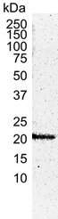
Western blot analysis of a HEK293 cell lysate with murine anti-human H-Ras antibody (MCA2884) followed by detection with a goat anti-mouse HRP conjugated secondary antibody. Visualization was performed by Chemiluminescence.
Featured Murine Anti-H-Ras Antibody (MCA2884)
The Ras protein family encompasses three members HRAS, NRAS and KRAS.
The genes encoding these proteins present some of the best studied proto-oncogenes, which upon mutation are involved in inducing various cancer types (including bladder, cervical, pancreatic and prostate).
Murine anti-human H-Ras antibody (MCA2884) is frequently used in Western blotting experiments and has been successfully used for the detection of H-Ras in human epitheliod breast cancer cells (MCF10A) (Vuoriluoto et al. 2011).
Applications: ELISA and Western Blotting
Citations: 2
View all Antibodies for the Analysis and Characterization of Oncoproteins Cell Proliferation Reagents
References
- Lodish H et al. (2000). Molecular Cell Biology, 4th edition. New York: W. H. Freeman; 2000.
- Condon LT et al. (2002). Overexpression of Bcl-2 in squamous cell carcinoma of the larynx: a marker of radioresistance. Int J Cancer. 2002 Aug 1;100(4):472-5.
- Vuoriluoto K et al. (2011). Vimentin regulates EMT induction by Slug and oncogenic H-Ras and migration by governing Axl expression in breast cancer. Oncogene. 2011 Mar 24;30(12):1436-48.